Nanotechnology Stock Images
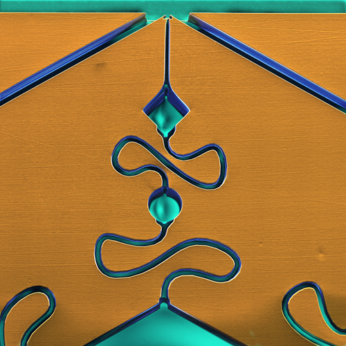
BC5084 - Rennet, stomach lining of a ruminant, on a chip. Micro-Electro-Mechanical Systems (MEMS) provide complete systems-on-a-chip, by providing sense and control of the environment for microelectronic integrated circuits. In this case, MEMS control rennet functions, such as transportation of liquids through capillary-strengths. © Science Source - science photos
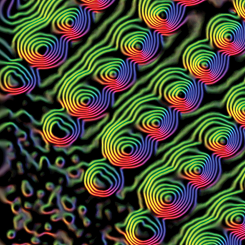
BU9846 - This image of magnetic flux lines around individual nickel nanodots reveals the local distribution of electromagnetic potential. ©Science Source - science photos
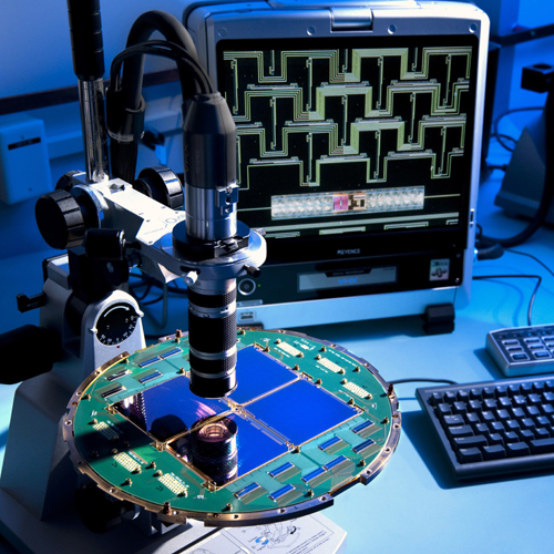
JA1756 - Devices that use superconductivity to gather, filter, detect, and amplify polarized light from the cosmic microwave background (relic radiation left over from the Big Bang) for NASA's BICEP2 telescope at the South Pole. © Science Source - science photos
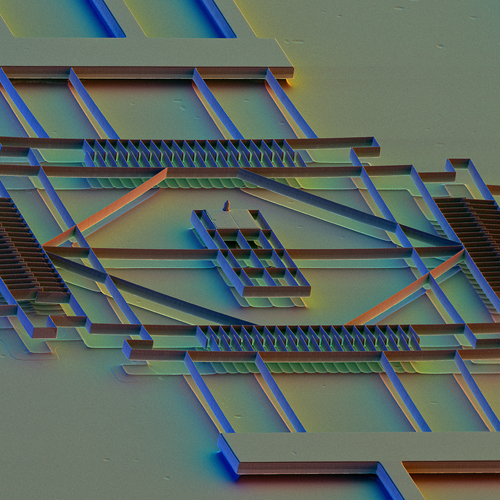
JA5154 - Color-enhanced Scanning Electron Micrograph showing a MEMS (microelectromechanical systems) Scanning Tunneling Microscope (STM). © Science Source - science photos

JA5685 - Color-enhanced Scanning Electron Micrograph (SEM) of nano-fabricated nichrome micro-wires; made at U.C.L.A. © Science Source - photos
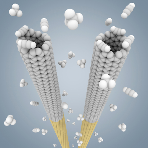
JC2670 - Cloning nanotubes: In this computer model, small, pre-selected nanotube "seeds" (yellow) are grown to long nanotubes of the same twist or "chirality" in a high-temperature gas of small carbon compounds. © Science Source - science source
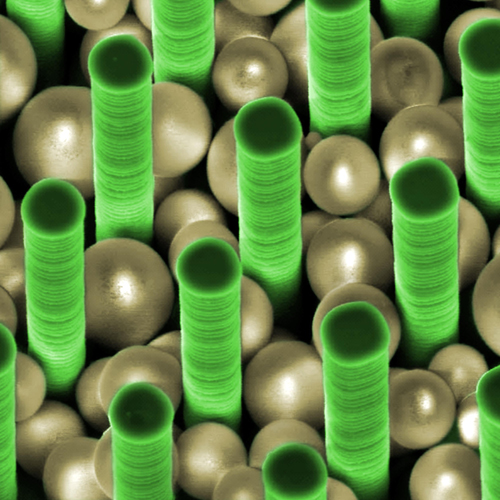
JC2889 - Ordered pillar arrays have been successfully explored as advanced porous media for separations. As a logical extension of this approach and to increase the surface area, silicon pillar arrays with embedded silica nanospheres have been implemented.
© Science Source - science photos

JC2890 - Silicon nanowires grown on a patterned substrate - a model system of different materials nanowire growth that will be used as advanced media for photovoltaics and energy storage applications.
© Science Source - science photos
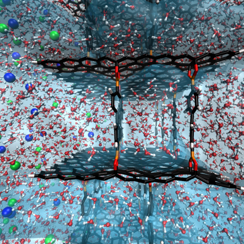
JC2899 - Simulations by Oak Ridge National Laboratory and Rensselaer Polytechnic Institute reveal the potential of graphene oxide frameworks, pictured in black, to remove contaminants such as salt ions, seen in blue and green, from water.
© Science Source - science photos

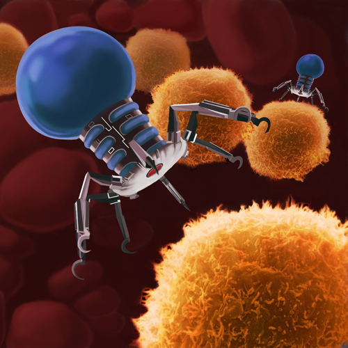
JA0988 - Medical nanorobot aiding red blood cell. © Science Source - science - photos
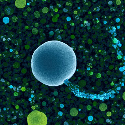
JC3975 - SEM showing tiny silicon nanostrands (green) and balloons and speckles of indium (blue). © Science Source - science photos
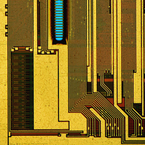
JC3708 - Transmitted light micrograph of a synchronous dynamic random-access memory (SDRAM) module (capacity 128 MB, PC-100 series). © Science Source - science photos

JC3993 - Graphene, a layer of carbon just a single atom thick, forms crystals when grown on copper. This "snowflake" is about 75 microns in size––about the diameter of a single human hair. © Science Source - science photos
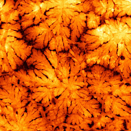
JC4311 - What appear to be lovely autumn leaves are actually the dendritic sprawl of lithium growing inside a battery, imaged via transmission electron microscopy. © Science Source - science photos
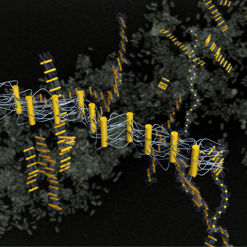
JC4316 - Nanorods form "rungs" on ladder-like ribbons linked by multiple DNA strands in a unique arrangement resulting from the collective interactions of the flexible DNA tethers. © Science Source - science photos
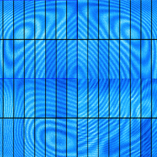
JC4319 - These digital camera CCDs (charge-coupled devices), totalling four chips with 16 segments each, will join 196 others inside the 3.2-gigapixel camera of the Large Synoptic Survey Telescope (LSST). The false-color image shows the CCD chips illuminated by near infrared light, which is the wavelength range where the LSST will study distant galaxies in its quest to reveal the secrets of dark energy and dark matter. The patterns are interference fringes caused by small thickness variations in the silicon wafer from which they were fabricated, but these striking contours will not impact performance.
© Science Source - science photos
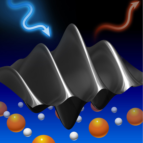
JC4320 - In this rendering, never-before-seen magnetic excitations ripple through a high-temperature superconductor, revealed for the first time by the Resonant Inelastic X-ray Scattering technique. By measuring the precise energy change of beams of incident x-rays (blue arrow) as they struck these quantum ripples and bounced off (red arrow), scientists discovered excitations present throughout the entire LSCO phase diagram. © Science Source - science photos
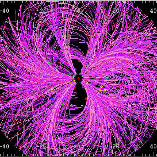
JC4321 - Event display of a single collision of 200 GeV gold ions as measured by the silicon vertex tracker at the Relativistic Heavy Ion Collider's PHENIX detector. © Science Source - science photos
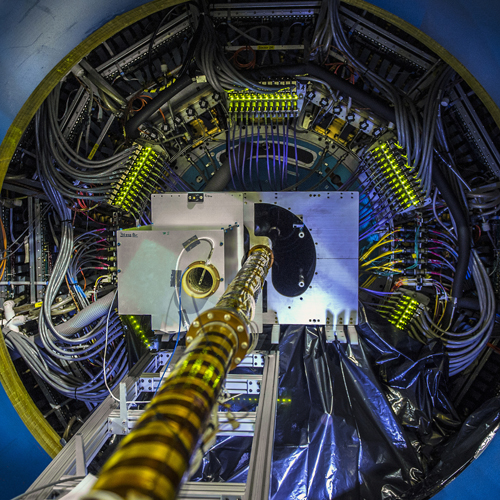
JC4329 - Shown here is the central portion of the Heavy Flavor Tracker (HFT) being installed at the Relativistic Heavy Ion Collider's STAR detector. The HFT will track particles made of "charm" and "beauty" quarks, rare varieties or flavors that are more massive than the lighter "up" and "down" quarks that make up ordinary matter. © Science Source - science photos

RD0449 - Nanotechnology research, conceptual computer artwork. © Science Source - science photos

RD0478 - Super buckyball molecule, computer artwork. © Science Source - science photos

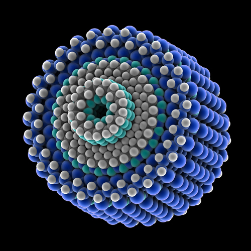
RD0514 - Medical nanoparticles, conceptual computer artwork. © Science Source - science photos
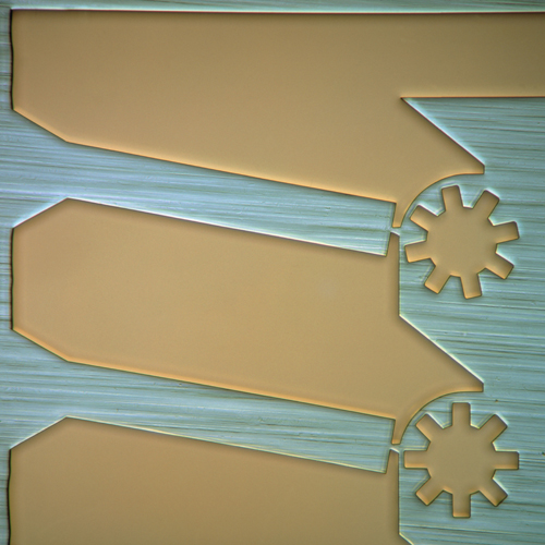
SN9674 - Light micrograph of components of a micromechanic pump. These were made using similar techniques to those employed in integrated circuit (silicon chip) manufacture. © Science Source - science photos


