Medical Stock Photography & Stock Medical Illustration Gallery
Buy Medical Illustration and Medical Photo Framed Prints and Posters
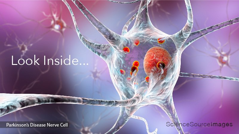
Parkinson's disease nerve cells. Computer illustration of human nerve cells affected by Lewy bodies (small red spheres inside cytoplasm of neurons) in the brain of a patient with Parkinson's disease. Lewy bodies are abnormal accumulations of protein that develop inside nerve cells in Parkinson's disease, Lewy Body Dementia, and some other neurological disorders.

An anterior view of the right breast. Illustration dipicting cancer in glandular tissue and lymph nodes of the breast.

Genetic research. Pipette adding a sample to a petri dish with a DNA (deoxyribonucleic acid) gel in the background.

Anterolateral view of a sectioned eye. The retina, the layer of tissue on the inner wall of the eye, contains specialized cells called rods and cones that are responsible for the sensation of light.

Woman with a medical patch. Patches that adhere to the skin and release drugs or hormones are now available for nicotine withdrawal, birth control, weight loss, and depression.
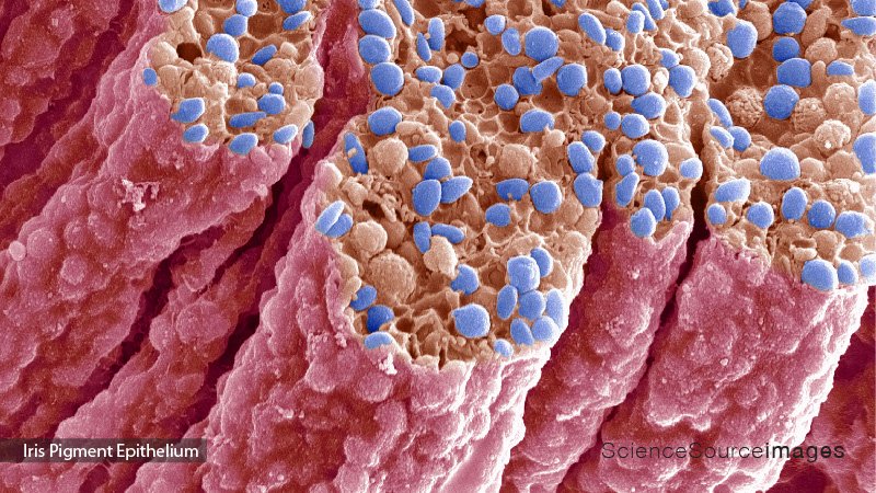
Iris pigment epithelium. Colored scanning electron micrograph (SEM) of a section through the iris of an eye, showing the iris pigment epithelium (IPE).

Psoriasis, a disease which affects the skin and joints. Psoriasis causes excessive skin production, resulting in red scaly patches and inflammation.

Arthritic hands. Colored X-ray of the deformed hands of a patient suffering from rheumatoid arthritis. The patient's fingers are abnormally bent because of damage to the joints (red). Rheumatoid arthritis is an autoimmune disorder.

Spring flowers representing human lungs, conceptual studio shot.

Anthrax bacteria, computer illustration. Anthrax bacteria (Bacillus anthracis) are the cause of the disease anthrax in humans and livestock.
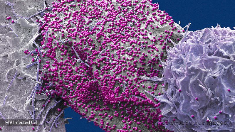
HIV infected 293T cell. Colored scanning electron micrograph (SEM) of a 293T cell infected with the human immunodeficiency virus (HIV, pink dots).
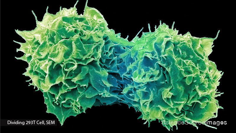
Dividing 293T cells in culture, colored scanning electron micrograph (SEM). These cells were isolated from human embryonic kidneys in 1977 and have been growing in culture ever since.

Parkinson's disease. Computer artwork of neurons (nerve cells, pink) containing Lewy bodies (green). Lewy bodies, which are deposits of protein, are found in neurons in the brains of patients with Parkinson's disease. It is thought that they cause the progressive degeneration of the neurons that leads to the symptoms of Parkinson's.
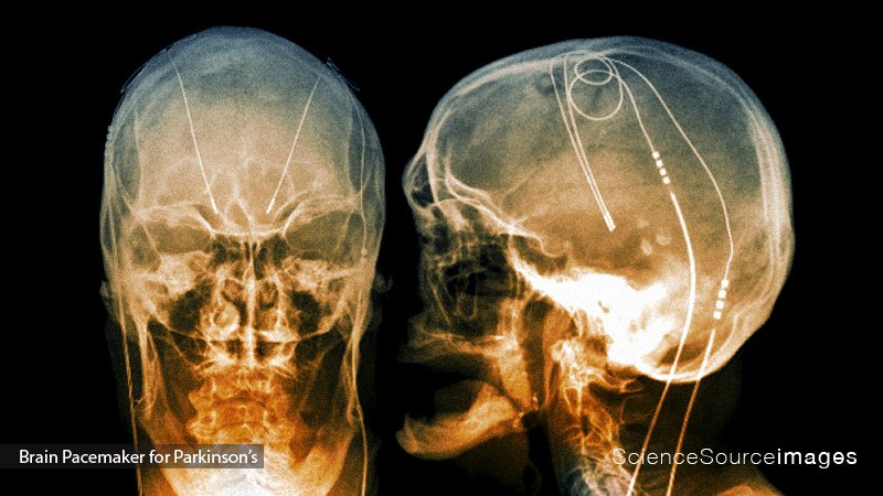
Parkinson's brain pacemaker. Coloured X-rays of sections through the head of a 61-year-old patient with Parkinson's disease (PD), showing the electrodes (light lines) of a deep brain stimulator (DBS) implanted in the brain.
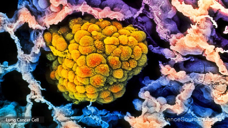
Lung cancer. Colored scanning electron micrograph (SEM) of a small cancerous tumor (orange) filling an alveolus of the human lung.

The human fetus after 10 weeks shows the legs & feet (note development of the toes), & the spiraling umbilical cord carrying the blood vessels which exchange nourishment (ie oxygen) & waste products with the maternal circulation.
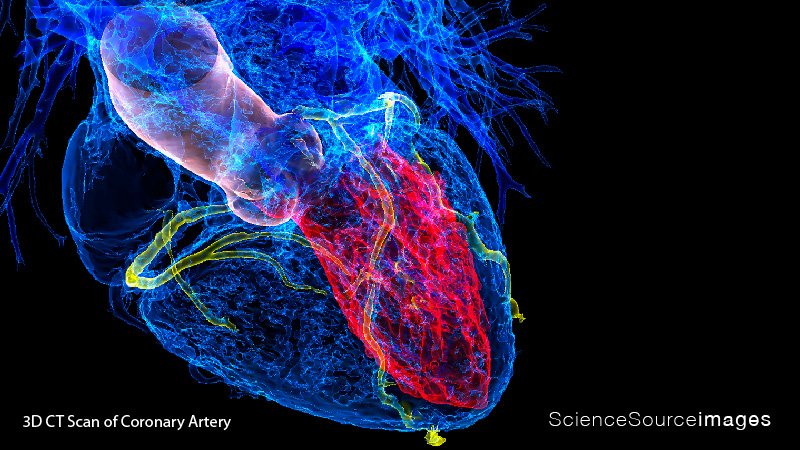
Heart in coronary artery disease. Illustration based on a 3D computed tomography (CT) scan of a human heart affected by coronary artery disease (CAD).
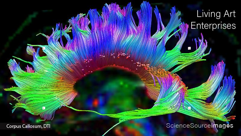
Corpus Callosum Tractography generated as strings. Diffusion tensor imaging (DTI) with generation of color diffusion tractography can have significant impact on how a brain tumor is resected.

Woman signing the word "Leaf" in American Sign Language while communicating with her son.
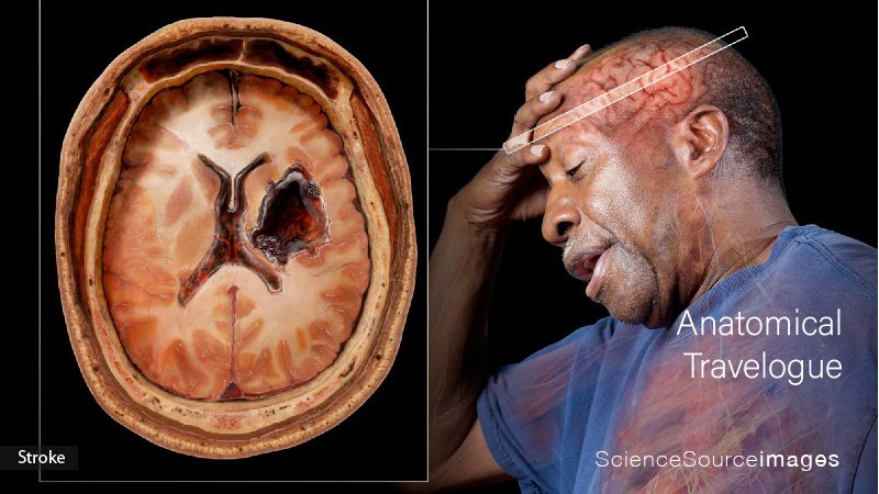
Medical visualization taken from human scanned data showing a man experiencing a stroke. Strokes occur when blood supply to the brain is cut off.


